Lipemia: The Optical Illusion That Breaks Assays
Fat in the blood doesn't just look bad; it scatters light and clogs filters. How lipemia distorts cfDNA values and how to handle the 'milky' sample.
Lipemia: The Optical Illusion That Breaks Assays
We love our Schnauzers, Miniature Pinschers, and other breeds prone to hyperlipidemia. What we don't love is their milky, lipemic blood samples.
Lipemia—elevated triglycerides and/or cholesterol causing visible cloudiness in plasma—is a common pre-analytical challenge in veterinary medicine. For standard chemistry panels, most modern analyzers can compensate for mild to moderate lipemia. But for cfDNA testing, lipemia is a major interference that can produce unreliable results through multiple mechanisms.
Understanding why lipemia is problematic, and how to manage it, is essential for any practice offering liquid biopsy services.
What Causes Lipemia?
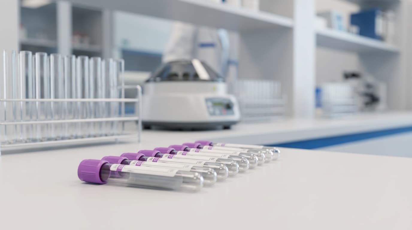
Lipemia results from elevated lipoproteins in the blood, causing the plasma to appear turbid, milky, or opaque instead of its normal clear, straw-yellow color.
Common Causes
Post-Prandial Lipemia (Most Common):
- Drawing blood shortly after a meal, especially a fatty meal
- Triglycerides peak 3-6 hours after eating
- Usually clears by 8-12 hours post-meal
Pathological Hyperlipidemia:
- Hypothyroidism (common in dogs)
- Diabetes mellitus
- Cushing's disease
- Pancreatitis
- Primary idiopathic hyperlipidemia (common in Miniature Schnauzers)
- Protein-losing nephropathy
- Cholestasis
Breed Predispositions:
- Miniature Schnauzers: Primary hypertriglyceridemia
- Shetland Sheepdogs: Familial hypercholesterolemia
- Beagles: Various lipid abnormalities
- Any breed with underlying endocrine disease
How cfDNA Is Measured: The Fluorometry Principle
To understand why lipemia interferes, we need to understand how cfDNA concentration is typically measured.
The Fluorometric Method
Most laboratories use fluorometry-based methods to quantify cfDNA:
1. The Dye: A fluorescent dye (such as PicoGreen, SYBR Gold, or Qubit reagents) is added to the sample
2. The Binding: The dye molecules insert themselves between DNA base pairs (intercalation)
3. The Excitation: A light source shines on the sample at a specific wavelength
4. The Emission: Bound dye molecules emit light (fluoresce) at a different wavelength
5. The Measurement: A detector measures the intensity of the emitted light
6. The Calculation: Fluorescence intensity is proportional to DNA concentration; the result is calculated from a standard curve
This method is elegant, sensitive, and works beautifully—for clear samples.
How Lipemia Breaks the System
Lipid droplets suspended in plasma interfere with fluorometry through multiple mechanisms:
1. Light Scattering
Lipid droplets scatter light in all directions:
- Excitation scattering: The excitation light scatters before reaching all the DNA-dye complexes
- Emission scattering: The fluorescent signal scatters instead of reaching the detector directly
- Rayleigh scattering: Small lipid particles scatter light proportional to the fourth power of frequency, creating complex optical effects
Result: Unpredictable—can cause either artificially high or artificially low readings depending on the specific optical geometry.
2. Quenching (False Low)
The lipid droplets can physically block light pathways:
- Excitation light doesn't penetrate the sample fully
- Emitted fluorescence is absorbed or scattered before reaching the detector
- The sample appears to have less DNA than it actually does
Result: False negative or underestimated cfDNA concentration.
3. Autofluorescence (False High)
Certain lipid-protein complexes can emit their own fluorescence:
- Lipofuscin (a lipid-protein aggregate) naturally fluoresces
- Some lipoproteins have intrinsic fluorescence
- This background fluorescence adds to the DNA signal
Result: False positive or overestimated cfDNA concentration.
4. Variable Interference
The worst aspect of lipemia interference is its unpredictability:
- The direction of error (high vs. low) depends on the degree of lipemia, the specific wavelengths used, and the optical path
- Mild lipemia might scatter light (false low)
- Different lipemia might contribute autofluorescence (false high)
- You cannot reliably predict or correct for the error
Impact on DNA Extraction
Beyond measurement interference, lipemia affects sample processing:
Spin Column Clogging
Many DNA extraction protocols use spin columns—small tubes with silica membranes that bind DNA:
1. Sample is loaded onto the column
2. DNA binds to the silica membrane
3. Washes remove contaminants
4. DNA is eluted
The Problem: Lipids can clog the silica membrane, preventing proper binding and washing. DNA may be lost during the extraction process.
Result: Falsely low cfDNA concentration due to extraction failure, not measurement error.
Reduced Extraction Efficiency
Even when columns don't fully clog:
- Lipid coating on the membrane reduces DNA binding capacity
- Lipids may elute with DNA, carrying interference into the final sample
- Reproducibility of extraction decreases
Managing Lipemic Samples
Prevention: The Best Strategy
Fast the Patient:
- A 6-hour fast clears most post-prandial lipemia
- 8-12 hours is even better for dogs with borderline lipemia
- Water should remain available (no fluid restriction)
Schedule Strategically:
- Morning appointments after overnight fast are ideal
- Avoid drawing immediately after meals
Know Your High-Risk Patients:
- Miniature Schnauzers and other predisposed breeds may need longer fasts
- Patients with diabetes, hypothyroidism, or Cushing's may have persistent lipemia
- Repeat fasted draws if initial sample is lipemic
Processing: Removing Lipid Contamination
The double-spin protocol can help remove some lipid content:
High-Speed Spin Separation:
When you centrifuge plasma at high speed (16,000 x g), lipids stratify based on their density:
- Top layer: White, creamy lipid layer (chylomicrons, VLDL)
- Middle: Clearer plasma
- Bottom: Any remaining cellular debris
Technique:
1. Perform the standard double-spin protocol
2. After the second spin, observe the tube carefully
3. If a lipid layer is visible at the top:
- Use a clean pipette to carefully aspirate the lipid layer off the top, OR
- Insert the pipette through the lipid layer and withdraw plasma from the middle, leaving both top (lipid) and bottom (debris) behind
4. Transfer clear plasma to final tube
Limitations:
This technique works for chylomicrons and large lipid particles. Smaller lipoproteins (LDL, HDL) remain suspended in solution and cannot be removed by centrifugation.
When to Reject and Redraw
Some samples are simply too lipemic to salvage:
Reject if:
- Plasma is opaque (cannot see through it)
- Lipid layer reforms after removal attempts
- Patient has known severe hyperlipidemia
Action:
- Document the rejection and reason
- Educate the owner about fasting requirements
- Reschedule with appropriate preparation
- If pathological hyperlipidemia is suspected, consider addressing the underlying cause before attempting liquid biopsy
Lipemia Grading Scale
Many labs use a visual or spectrophotometric lipemia index:
| Grade | Appearance | cfDNA Testing Decision |
|-------|------------|------------------------|
| 0 | Clear, transparent | ✅ Proceed |
| 1+ | Slightly hazy | ⚠️ Proceed with caution; note in records |
| 2+ | Moderately turbid | ⚠️ Attempt lipid removal; may need to reject |
| 3+ | Milky, opaque | ❌ Reject—redraw after fasting |
Special Considerations
Diabetic Patients
Dogs with uncontrolled diabetes may have persistent hypertriglyceridemia:
- Even fasted samples may be lipemic
- Consider glycemic stabilization before elective liquid biopsy screening
- For urgent testing, document the lipemia and interpret results cautiously
Miniature Schnauzers
This breed deserves special mention due to their high prevalence of idiopathic hypertriglyceridemia:
- May have lipemic samples even when fasted
- Chronic lipemia may be their baseline
- Consider whether liquid biopsy is the right test, or whether alternative approaches are needed
- If proceeding, document extensively and interpret with caution
Post-Prandial Emergency Cases
In emergency situations where a patient presents non-fasted:
- Prioritize clinical care over ideal sample collection
- If cfDNA testing is urgent, document the fed status
- Consider whether the information is still valuable despite potential interference
- Plan for confirmatory testing once the patient is stabilized and can be fasted
Summary: The Clear Sample Standard
| Factor | Best Practice |
|--------|---------------|
| Patient preparation | 6-12 hour fast |
| Visual inspection | Check for turbidity before processing |
| Processing | High-speed spin can remove some lipids |
| Rejection threshold | Opaque samples should be rejected |
| Documentation | Note lipemia grade in records |
| Interpretation | Results from mildly lipemic samples require caution |
If the sample looks like milk, it's better to reschedule than to pay for an unreliable guess. Prevention through proper patient preparation is always the best strategy.